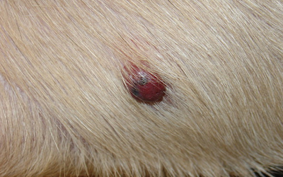
Hemangiosarcoma in Dogs. The lesions consisted of approximately 13 x 3 cm and 15 x 10 cm irregular patchy regions of 05-30 cm circular sometimes raised redd.

Hemangiosarcoma is a type of cancer that arises from blood vessels.
Hemangioma skin dogs. By the time a hemangiosarcoma is seen in the skin the cancer has usually spread to the dogs organs. A veterinarian will not be able to tell you if the growth is a hemangioma or a hemangiosarcoma just by examining it. A biopsy or at least a cytology is required.
To be completely sure which type of tumor you are dealing with a biopsy is best. What is Hemangioma in Dogs. This type of growth a cutaneous hemangioma is a neoplasm on the skin that is benign.
It resembles a blood blister or angiokeratoma. Hemangiomas are lesions of the vascular system that are formed by the cells that are responsible for forming blood vessels or endothelial cells. Dermal hemangiosarcoma tumors appear as red or sometimes black growths on your dogs skin.
This form of hemangiosarcoma is most often associated with exposure to the sun so it will more likely form on areas that are naked or lightly furred like the abdomen. Dogs with short white fur are most susceptible to dermal hemangiosarcoma. Hemangiosarcoma in Dogs.
Hemangiosarcoma is a type of cancer that arises from blood vessels. Hemangiosarcomas can develop on the skin under the skin in the subcutaneous tissue and on internal organs such as the spleen and heart. These are rare tumors in cats but common in dogs.
Skin had more hemangiomas and hemangiosarcomas of the dermis 65 than did dogs with variable length hair coats and pigmentation 28. Dogs with short hair coats and lightly pigmented skin had fewer hemangiomas and hemangiosarcomas of the subcutaneous tissue 10 than did dogs with variable length hair coats and pigmentation 22. Although the cause of vascular tumors in dogs and cats has not been determined there appears to be a greater incidence of hemangiosarcoma of the skin in lightly pigmented and sparsely haired dogs which may indicate that ultraviolet light sun exposure may play a role in the development of these tumors.
They are found in older dogs and cats and appear closer to the surface of the skin. In dogs these tumors are most commonly recognized in Peekapoos Old English Sheepdogs and English Springer Spaniels. They are often smaller firmer and less cystic than apocrine adenomas.
Hemangiomas in dogs are generally benign soft tissues and skin tumors while sarcomas are malignant tumors developing in the soft tissuesHemangiosarcoma or cancer of the blood vessels is a type of tumor occurring more often in canines than any other animalUnfortunately hemangiosarcoma symptoms often dont become obvious until the cancer has already spread. Hemangiosarcoma is a cancer of the blood vessel lining. Technically called the vascular endothelium.
Hemangiosarcoma can spread fast. Because your dog has blood vessels everywhere in his body. Its also called angiosarcoma or malignant hemangioendothelioma.
Hemangiosarcoma is abbreviated as HSA. In addition subcutaneous hemangiomas are typically elevated partially hairless and blue in color. Skin hemangiomas appear as a small dome with a reddish-black tint.
These tumors may be caused by certain chemicals the sun or they may be idiopathic unknown cause in origin. Cutaneous histiocytomas are a benign skin tumor of young dogs 1-3 years. A cutaneous hemangioma is a benign tumor found on the skin of dogs.
It originates either in the dermis or the subcutaneous layer of the dogs skin. Hemangiomas are formed by endothelial cells the cells that form blood vessels. Hemangiomas are small masses of blood vessels and are red to black in color.
The relationship between skin pigmentation and piliation and the development of hemangiomas and hemangiosarcomas in the dermis and subcutaneous tissue was studied in 212 dogs. These 212 dogs had a combined total of 306 tumors. 38 of these 212 dogs had two or more of the same tumor in a different loc.
A 1-year-old spayed female mixed-breed dog had two reddish-purple cutaneous lesions one on the right dorsal antebrachium and the other on the right shoulder. The lesions consisted of approximately 13 x 3 cm and 15 x 10 cm irregular patchy regions of 05-30 cm circular sometimes raised redd. JPC SYSTEMIC PATHOLOGY.
Unspecified breed and age dog HISTORY. A small dermal mass HISTOPATHOLOGIC DESCRIPTION. Haired skin and subcutis.
Expanding the dermis and subcutis elevating the mildly hyperplastic epidermis and compressing adnexa is a 5 X 10 mm. Ous skin of sparsely haired dogs9 Histologically endothelial tumors in animals are most commonly designated as angiomatosis he-mangiomas and hemangiosarcomas. Several var-iants of hemangiomas are described such as capillary and cavernous hemangiomas infiltrative hemangioma arteriovenous hemangioma gran-.
If its skin hemangiosarcoma the survival rate could be higher. Assess your dogs quality of life. Some skin hemangiosarcoma surgery can be successful and reduce the chances of HSA spreading internally.
However if you see weight loss lethargy lack of moment obvious pain and your dog not eating then euthanasia has to be a consideration. Hemangiosarcoma is Blood or Skin Cancer in Dogs and Cats Hemivertebrae are Congenitally Deformed Vertebra in Dogs and Cats Hepatic Encephalopathy in Dogs and Cats.