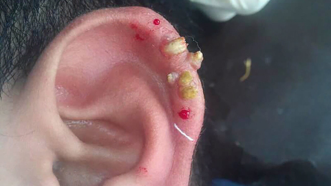
Less often cutaneous horns form in dogs as a result of a canine papillomavirus infection. If the fluid exits the horns and enters the vagina the dog will begin to groom herself excessively exposing bacteria to the vagina and cervix.

Carl pops a baseball size sebaceous cyst on a k9 patient.
Horn cyst dog. These cysts of which there are several kinds are filled with keratin which is a skin protein. Keratin is a structural protein that makes up hair horns yes rhino horn hooves and the outer layer of skin. Keratin protects epithelial cells which form the under layers of skin from damage.
Most cysts are hard or have a solid core. Less often cutaneous horns form in dogs as a result of a canine papillomavirus infection. Cornifying means looking like a horny substance and epitheliomas is a medical word for a skin tumor that can be either benign or cancerous.
A cutaneous horn on a dog will be a growth that sticks up from the skin surface. It can feel like a stick-like growth on a dogs tail. It appears as lumps seldom more than 04 inches 1 centimeter in diameter often with a shiny horn-like surface.
Among dogs Manchester Wheaten and Welsh Terriers are at greatest risk. The head and abdomen are affected most often. As the lining thickens from cystic endrometrial hyperplasia fluid builds up in the horns of the uterus causing them to swell.
The uterus expands becoming uncomfortable. If the fluid exits the horns and enters the vagina the dog will begin to groom herself excessively exposing bacteria to the vagina and cervix. This time though the open wound had become infected and needed a cleanup and antibiotics.
Cysts tend to occur in middle-aged or older dogs and are most commonly linked to breeds such as German Shepherds Boxers and Cocker Spaniels. Technically speaking a cyst is a sac in the skin lined with secretory cells. Dogs need to have their cysts checked out for the same reason that humans have moles and breast lumps checked out.
While the cysts could be benign theres always a chance that it could be something serious. Cysts can also interfere with a dogs normal movement and impair them. A cyst on a paw pad leg or joint can interfere with their ability to walk.
Skin cysts on dogs are sacs that are lined with secretory cells and which are found on the skin. Once the sacs form there is a buildup of fluid resulting in swollen bumps on the skin. While some of the cysts will retain the fluids others will dry out.
These masses may be found anywhere on the body. Sebaceous cysts are benign but can also be mistaken for a malignant tumor called a sebaceous gland adenocarcinoma or a benign mass called a sebaceous gland adenoma. If the cyst does not bother your dog your vet might leave it alone but a cyst can be surgically removed if necessary.
Interdigital cysts are a common recurrent and painful problem in many dogs. Treating the secondary infections will often result in only a short to medium term resolution. Recurrence is very likely unless the primary cause is detected treated.
The paw s may be inflamed only or inflamed and infected with any combination of microbes bacteria. Cutaneous horn cornu cutaneum is a projectile conical dense hyperkeratotic nodule that resembles the horn of an animal The horn is composed of compacted keratin. A number of skin lesions can be found at the base of this keratin mound.
Cutaneous horns most frequently occur in sites that are exposed to actinic radiation or burns and hence are typically. Cutaneous horns also known by the Latin name cornu cutaneum are unusual keratinous skin tumors with the appearance of horns or sometimes of wood or coral. Formally this is a clinical diagnosis for a conical projection above the surface of the skin They are usually small and localized but can in very rare cases be much larger.
Although often benign they can also be. A cutaneous horn is a hornlike growth 12 to 2 with unknown cause. It can be a byproduct of either follicular cysts or cancer.
Some dogs have an allergic reaction to the larva. Treatment involves surgical removal. Treatment involves opening the nodule and then removal of any larva.
This is a type of fly larva. A dog skin lump forms around the larva which. Dogs with Horners Syndrome show the following signs of the affected eye.
A constricted small pupil elevated third eyelid a drooping upper eyelid sunken appearance of the eye and dilation of the blood vessels on the affected side of the face which makes the area warmer and reddened. Hyperkeratosis is a skin condition that results from an overstimulation of keratin production in your dogs body. If youre unfamiliar with keratin its the same fibrous protein that our hair and nails are made of as well as hoofs and horns on other animals.
Carl pops a baseball size sebaceous cyst on a k9 patient. You will either find this extremely gross or extremely awesome if youre the latter- enjoy. Cyst wall commonly contains secondary horn cysts complex.
Skin Diseases of the Dog and Cat. Kok MK Chambers JK Ong SM Nakayama H Uchida K. Hierarchical Cluster Analysis of Cytokeratins and Stem Cell Expression Profiles of Canine Cutaneous Epithelial Tumors.
Papillomas or warts can turn into hard horny growths that are similiar in consistency to nails or a rhino horn. The growth may look like a cone or horn and it can vary in size. The name comes from the growth sometimes resembling an animals horn.
This skin condition is more common in older adults and. Follicular cysts are usually thin walled lined with a granulosa cell layer and contain a clear watery fluid. Thus they resemble Graafian follicles.
It is believed that follicular cysts arise because of a failure of luteinizing hormone LH release or a lack of response of a Graafian follicle to LH Lopate Foster 2010. Hair follicle tumors develop as a result of the disordered growth of the hair follicles. There is a genetic predisposition to developing hair follicle tumors and they are more likely to occur in the Golden Retriever Standard Poodle Miniature Schnauzer English Springer Spaniel Irish Setter German Shepherd Cocker Spaniel Norweigan Elkhound Keeshond Kerry Blue Bedlington.
A horn cyst is a large thin granular cell layer that has been completely keratinized without retained nuclei. There may be a difference between truth and pseudotruth. A pseudo horn cyst is a skin condition characterized by significant sectioning of the papillomatous.