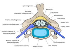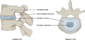
There are seven cervical vertebrae in every dog even if some dogs may have a longer neck than others. The dogs spine is divided into cervical vertebrae thoracic vertebrae lumbar vertebrae sacral vertebrae and coccygeal vertebrae.

The external anatomy of a dog is quite simple to understand.
Vertebrae anatomy dog. Thoracic vertebrae in dogs or canine. The thoracic division of dog spine anatomy consists of the thoracic vertebrae thick intervertebral discs spinal cord and thoracic spinal nerves. Now I will show you some unique anatomical facts of the thoracic vertebrae of a dog so that you may identify it so quickly.
The dogs spine is divided into cervical vertebrae thoracic vertebrae lumbar vertebrae sacral vertebrae and coccygeal vertebrae. There are seven cervical vertebrae in every dog even if some dogs may have a longer neck than others. Anatomy of the canine lumbar vertebrae and lumbosacral junction CT This anatomical module of the atlas of veterinary radiological anatomy vet-Anatomy covers the lumbar vertebrae lumbosacral joint sacrum and caudal vertebrae of the dog on a Computed Tomography CT and on 3D images of the lumbar spine and pelvic girdle.
From Evans HE. Millers anatomy of the dog ed 4 Philadelphia 2013 WB Saunders All vertebrae except the sacral vertebrae remain separate and form individual joints. Four sites with limited motion exist within the canine spine.
6 These sites occur at areas where the cranial and caudal articular surfaces are inclined in a nonparallel manner and in different. Speaking of skeletons a dog has 320 bones in their body depending on the length of their tail and around 700 muscles. Muscles attach to bones via tendons.
Depending on the breed of dog they will have different types of muscle fibers. Youve probably heard about slow and fast twitch muscle fibers before. Individually the joints between vertebrae are not very mobile the movement of the upper synovial joints is very limited just a sliding movement but as a whole the spine can perform movements of flexion and extension.
Intervertebral joints in dogs are like those of humans. Vertebrae are the bones of the spine. The spinal cord is the part of the CNS located within the vertebral canal.
It is involved in controlling movements of the trunk and limbs regulates visceral functions and transmits information to and from the brain. It extends from the foramen magnum in the occipital bone to the sixth or seventh lumbar vertebra in dogs. Costal cartilage 5 7.
This is an online quiz called Anatomy Canine Lumbar Vertebrae. There is a printable worksheet available for download here so you can take the quiz with pen and paper. Your Skills Rank.
This is an online quiz called Canine Vertebrae. There is a printable worksheet available for download here so you can take the quiz with pen and paper. Your Skills Rank.
You need to get 100 to score the 4 points available. The external anatomy of a dog is quite simple to understand. The following diagram and paragraph attempt to explain it in brief.
The muzzle is of varying lengths depending on the breed. Whiskers present on the muzzle are of some sensory use. Dogs also have a stop on their heads which is the point where the muzzle ends and the forehead begins.
Lumbosacral transitional vertebrae in dogs. Classification prevalence and association with sacroiliac morphology. The prevalence of lumbosacral transitional vertebrae LTV was determined by reviewing the pelvic radiographs of 4000 medium- and large-breed dogs of 144 breeds routinely screened for canine hip dysplasia.
They reach a maximum height a few vertebrae behind the cervicothoracic junction constituting the withers of the horse and then decline. The orientation of spinous processes shifts from caudo- to craniodorsal. The lumbar vertebrae are longer and more uniform in shape than the thoracic vertebrae.
Anatomy is at the core of the veterinary profession. Here we will begin to understand and learn the canine skeleton and key muscles which move it. Bones of the canine forelimb hindlimb and spine.
If we now move on to the spine of the canine. The canine spine is divided into five regions. Cervical thoracic lumbar sacral and caudal.
There are 7 cervical vertebrae 13 thoracic vertebrae 7 lumbar vertebrae and 3 sacral vertebrae. The number of caudal vertebrae varies according to the species. Cervical vertebrae edit edit source The atlas and axis are cervical vertebrae 1 and 2.
Cross-sectional anatomy of the canine thorax on CT imaging lungs trachea heart mediastinum diaphragma liver rib cage thoracic spine Dog - Pulmonary lobules - CT. Anatomy of the thorax of the dog on CT. Mediastinal vessels Aortic arch Mediastinum Heart Pulmonary arteries Pulmonary veins.
The canine spinal column is separated into four main regions. Cervical neck spine with seven vertebrae. Thoracic mid-back spine 13 vertebrae.
Lumbar lower back seven vertebrae. And sacral pelvic spine three vertebrae. It consists of sensory and sympathetic postganglionic fibers that innervate the dura mater the dorsal longitudinal ligament the vertebral venous sinuses and other vessels located in the vertebral canal Page 832 of Millers anatomy of the dog by Evans HE.
Computed tomographic anatomy of the canine cervical vertebral venous system. Computed tomographic CT venography of the cervical vertebral canal was performed in six clinically normal adult mixed-breed dogs from 14 to 23 kg. After dogs were euthanized and saline perfused a gelatin and iothalamate mixture was injected into the right external.
Canine Muscle Origins Insertions Actions and Nerve Innervations The purpose of this document is to provide students of canine anatomy a simple reference for muscular origins insertions actions and nerve innervations without having to search through the overwhelming verbiage that accompanies most canine anatomy texts. Thoracic Vertebrae and Ribs Fig. 8B-4 HORSES typically have a total of 18 thoracic vertebrae and 18 sets of ribs while CATTLE have a total of 13 thoracic vertebrae and 13 sets of ribs.
Observe the thoracic vertebrae ribs and costovertebral articulations on a skeleton. The vertebral extremity of the dog ribs possesses a round head a distinct neck and a tubercle. In the head of the dog ribs you will find the cranial and caudal articular facets.
These facets articulate with the socket that forms by the cranial and caudal costal fovea on the bodies of the two adjacent vertebrae.